

Cytopathic Effects in Uninoculated Cultures
Cytopathic Effects in Uninoculated Cultures
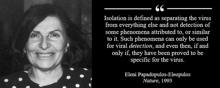
Control experiments invalidating virological methodology

Virology’s Lack of Control is an excellent article about the presence of cytopathic effects in uninoculated cultures.
Mike Stone outlines how the cell culture method used in virology was disproved in 1954 when John Enders observed cytopathic effects in uninoculated cultures.
This was confirmed by various other researchers over the next few years.
Some people might defend John Enders by claiming he could diffirentiate the cytopathic effects in the cultures because “internuclear changes typical of the measles agents” was not observed in the uninoculated culture.
However, such intranuclear changes do occur in uninoculated cultures aswell. To the surprise of virologists.
“To our surprise, measles virus intranuclear and intracytoplasmic eosinophilic inclusions occurred in both inoculated and uninoculated control HEK cultures. Thus, the adenovirus stock derived from the commercially made HEK cultures was inadvertently contaminated with a measles virus.”
Without any evidence, the authors claim that the uninoculated culture must have been “contaminated” with a measles virus.
Convenient rescue device highlighting the pseudoscience of virology.
Monkeys were not the only species whose kidneys were prone to spontaneous disintegration. In a 1970 study, it was reported that uninoculated goat kidneys were frequently breaking down.
“We found that goat kidney cells were also highly susceptible to measles virus, but uninoculated cultures also developed cytopathic effects frequently.”
Chimpanzee kidneys suffered the same fate in a 1963 study called Chimpanzee Kidney Tissue for Growth and Isolation of Viruses.
“During prolonged incubation (2 weeks or more) of uninoculated chimpanzee kidney tissue cultures, the cells frequently exhibited changes similar to changes caused by the growth of viruses.”
The authors suspected “adventitious viruses” and not the experimental procedure itself.
“The presence of adventitious viruses in some uninoculated chimpanzee kidney tissue cultures is suspected.”
This idea is unfounded, especially since innocous substances have been shown to induce cytopathic effects.
In a study published in 1962 it was found that yeast extract increases cytopathic effects and causes degenerative changes in both uninoculated and inoculated cultures.
“Experiments with yeast extract. Wittler, Cary, and Lindberg (1956) reported that yeast extract speeds up the growth of PPLO in tissue cultures and increases the cytopathic effect. When yeast extract (Difco) at a concentration of 0.5% was present in our cultures at the time of inoculation, both the control and the inoculated cultures showed degenerative changes within 1 week, probably because of toxicity of the extract for the cells.”
 – Welgene")
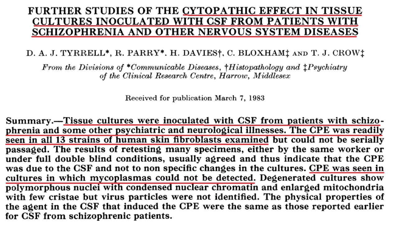
Fluids from people with “non-infectious” diseases have also shown to induce cytopathic effects.
“We have found that CSFs from patients with certain psychiatric syndromes, including schizophrenia, produce a cytopathic effect (CPE) when inoculated into stationary tissue cultures .. This CPE resembles that produced by certain viruses but no cytopathic agent has yet been established in tissue culture”
A study published in 1984 attempted to search for a viral etiology of inflammatory bowel disease. Cell cultures were used, but the authors concluded that the cytopathic effects observed were probably caused by non-replicating cytotoxic factor.
“Intestinal tissue filtrates induce cytopathic effects in inoculated cell cultures, but the effect we observed is non-specific … Our results suggest that the observed cytopathic effect was caused by a non-replicating cytotoxic factor.”
A 1970 publication demonstrated a clear indication that the experimental procedure itself create the cytopathic effects.
Kidney cells from healthy puppies were cultivated without the addition of an alleged “infected” sample. Yet, cytopathic effects were observed.
“Puppies serving as donors of kidney tissues for the cell cultures were derived from a closed colony of apparently healthy beagle dogs. The primary canine kidney cells were placed on maintenance medium consisting of Eagle’s minimal essential medium plus 2% fetal calf serum on the day after receipt of the monolayer cultures. Foci of CPE consisting of rounded cells and plaques (Fig. IA) appeared at 14 days after initiation of the cultures. The CPE progressed to involve 50 to 75% of the cell sheet by the end of the third week of cultivation.”

When virologists uninoculated control cultures exhibit cytopathic effects, they often blame “viral” contamination and not the experimental procedure itself. This can be seen in a 1970 study were an “adventitious virus” was blamed when the uninoculated cultures exhibited CPE.
“While attempting to isolate bovine rhinoviruses in BEK cell cultures, occasional controls were noted to degenerate spontaneously. The BEK cultures were incubated in roller drums at 33 C for isolation of bovine rhinoviruses. The maintenance medium contained Eagle basal medium with Earle salts, 2% normal rabbit serum, and the usual concentrations of antibiotics. On primary isolation, occasional uninoculated control cultures showed focal cytopathic effect (CPE) after 12 to 14 days of incubation.”
The idea of “endogenous viral contamination” goes back to 1955 when Rustigian et al. observed cytopathic effects in uninoculated cultures. Since then, virologists have been claiming to discover many viruses when their uninoculated cultures exhibit cytopathic effects.
“In recent years, there have been increasing numbers of reports concerning the recognition of latent virus infections in tissues of primates as well as nonprimates … As a result of the extensive use of primate cell cultures, a great number of simian viruses have been recovered from a variety of monkeys, baboons, and marmosets.
[…]
“The isolation of virus-like agents from monkey kidney tissue cultures was first reported by Rustigian et al. in 1955 (89). Subsequently, as a result of the extensive use of monkey kidney cell cultures, especially in the preparation of virus vaccines, a great number of simian viruses have been recovered as endogenous tissue contaminants by Hull and associates (56- 58).”
[…]
“On the basis of certain biological properties, especially cytopathic effect (CPE), simian viruses were originally divided into four groups by Hull et al. (57). Other biological properties, including plaque morphology, host-cell susceptibility, and hemagglutinin production, have also been used for grouping these viruses.”
[…]
“Recognition and characterization of simian viruses in cell cultures are of practical importance, since monkey tissues often harbor a variety of viruses.”
This convenient rescue device of endogenous viral presence have frequently been used by virologists in order justify their pseudoscientific methodologies. In his book Contamination in Tissue Culture, Jørgen Fogh outlines the widespread usage of this rescue device.
“Very many animal species have hidden virus infections, that is, viruses that appear to exist in close association with particular tissues or organs of the species involved, without causing overt symptoms. It follows that primary cultures prepared from almost any tissue may be found infected with viruses in vitro simply because the organ from which they were prepared was infected in vivo (Hsiung, 1968). In this way, primary cultures from chickens (Burke et al, 1965), chimpanzees (Rogers et al., 1967), dogs (Smith et al., 1970), ducks (Luginbuhl, 1968), guinea pigs (Hsiung and Kaplow, 1969), hamsters (Stenback et al., 1966), horses (Hsiung et al., 1969), man (Hsiung, 1968), rabbits (Nesburn, 1969), rats (Melendez et al., 1967), monkeys (Hsiung, 1968), and swine (Huygelen and Peetermans, 1967) have all been found to yield viruses spontaneously.”
When virologists don’t see the results they want, they often play around and add reagents until they do. In the case of the polio virus in Vero cells, scientists weren’t able to see the CPE they wanted until they added Trypsin. Of course, they blame it on “virus.”
“When monkey kidneys are infected in vivo with polio virus, no cytopathic effect can be observed and no virus can be recovered following subsequent perfusion of the kidney or homogenization of its tissue. However, if parts of an infected kidney found negative for virus by such tests are reduced to cell suspensions by trypsin treatment and used to set up conventional monolayer cultures, cell destruction eventually takes place, accompanied by the appearance of fully infective virus. When the kidney cells are removed from their in vivo situation to an alien environment, they somehow acquire susceptibility to polio virus.”
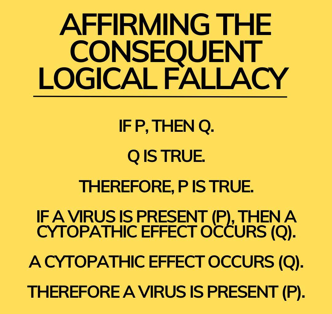
The presence of a viral agent in cell cultures was detected through the observation of cytopathic effects, or in the case of certain viruses, the detection of hemadsorption (the adherence of red blood cells to the surface of something). However, hemadsorption also occures in uninoculated cultures, as described by a 1961 publication.
“The presence of a viral agent in inoeulated cell cultures may be detected either by observing the cytopathic effect, i.e., cellular degeneration, in the inoculated cultures or, in the case of certain myxoviruses which do not give rise to distinct cellular degeneration, by the addition of red blood cells to the cell sheet and observance of hemadsorption. It is necessary to distinguish between cellular destruction due to specific viral effect and that due to toxicity of the inoculum … In employing hemadsorption as an indicator of the presence of viral agents, it must be kept in mind that certain simian viruses occur as natural contaminants of monkey kidney cell cultures, and that these may cause hemadsorption.”
The identification of the specific ‘viral agent’ was through the use of neutralization tests. The cell culture breakdown product was mixed with different protein brews and then added to cell cultures, and depending on what results happened to be seen in the cell cultures, a virus was determined.
“Identification of viral agents recovered in tissue culture systems is usually accomplished by the neutralization test. For this procedure, a standard amount of the unknown virus, usually 100 TCD50, is mixed with known immune serums, and after a suitable incubation period the serum-virus mixtures are inoculated into cell cultures in order to determine which, if any, of the immune serums is able to prevent cytopathogenesis or hemadsorption. A modification of the neutralization test which has proved particularly useful for the identification of viral agents is the metabolic inhibition test in which multiplication or nonmultiplication of the virus is detected colorimetrically. If the virus is specifically neutralized by an immune serum, cellular metabolism is unimpaired, and the medium is converted from alkaline to acid, as indicated by a shift in color of the phenol red indicator from red to yellow. For this procedure, the cells, virus and serum are all added to the test on the same day, eliminating the need for preliminary preparation of monolayer cell cultures in tubes.”
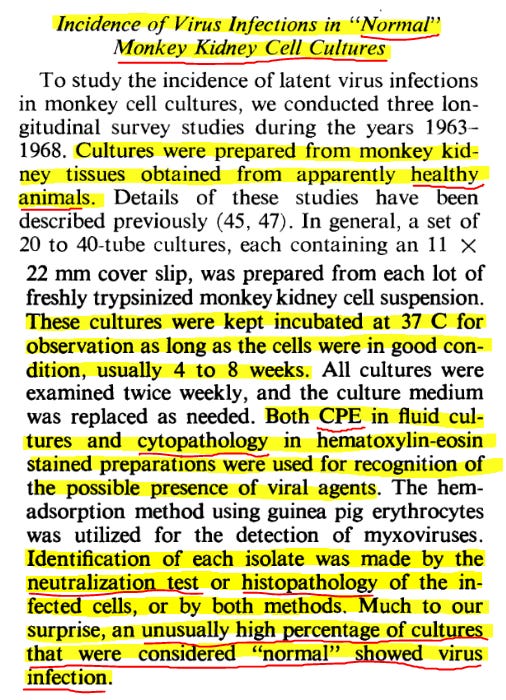
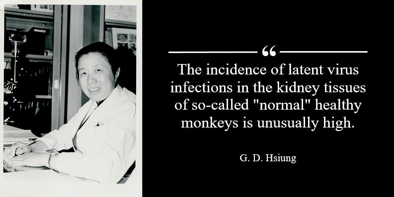
However, this methodology has been falsified due to control studies. In a 1968 publication entitled Latent virus infections in primate tissues with special reference to simian viruses, G. D. Hsiung et al. tested the surrogate markers, i.e. CPE and neutralization tests, on uninoculated normal cell cultures.
To their surprise, positive results from their surrogate markers were frequently observed in normal uninoculated cells.
“Much to our surprise, an unusually high percentage of cultures that were considered “normal” showed virus infection.”
The same effects attributed to viruses is seen in cell cultures even though no assumed infected material is added.
Typical distraction from the real cause of disease.
That's why they find the sequences and antibodies in healthy people.
But the sick people get the label they pre defined in order to cover up for the real causes.
Example: polio blamed for paralysis caused by ddt and arsenic based pesticides .
Great work Vil 💪💪