Particles Indistinguishable from Viruses
When imagined viruses turns out to be non-specific fragments of debris.
The field of virology was invented before establishing if viruses actually existed. There was no understanding how these hypothesized entities actually looked and functioned.
The existence of viruses were postulated on theoretical grounds, based on pathological changes in laboratory animals, induced by uncontrolled inoculations of filtered biological material. This was as noted by virologist Charles E. Simons in 1926.
“…he is inclined to look upon these smallest forms, which may properly be termed ultraviruses, as representing living matter of such intermediate type, the existence of which we have postulated on theoretical grounds.”
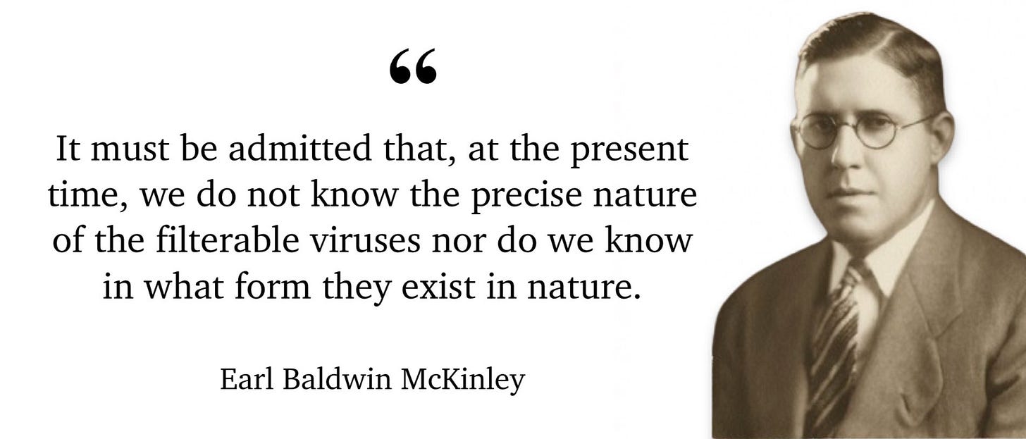
Virologist Karl F. Meyer expressed the uncertainty of the virus theory in a 1939 radio discussion.
“Doctor Meyer, will you summarize the present status of poliomyelitis in the field of experimental research? Dr. Meyer: That is a difficult question to answer directly, since we are still baffled by the simple question, “What is the disease agent in infantile paralysis?” … We do not know whether it is a living germ or something else growing in the cells of the brain. We cannot isolate it like a germ in a test tube, and it is too small to be seen with a microscope. There is only one species of animal—the Old World monkey—in which we can induce the same disease as seen in children. Yet these monkeys are not as susceptible as man, since there is no record of one monkey catching the infection from another monkey by exposure.”
In the 1940s, when the electron microscope was developed for biological material and commercial use, virologists finally had access to a powerful microscope to view their invisible entities.
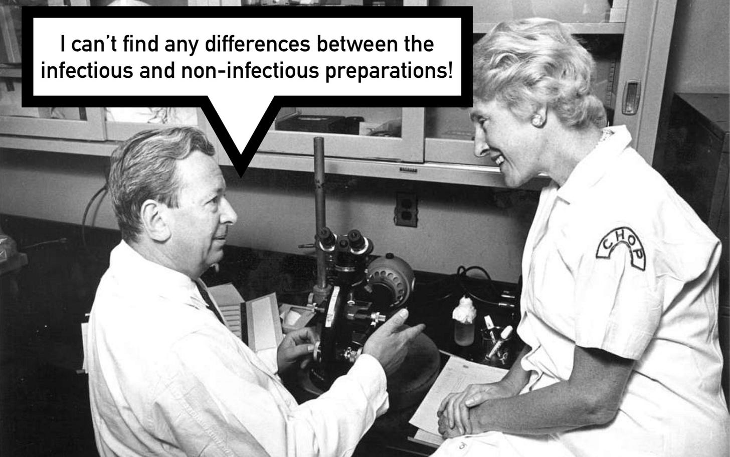
However, virologists could not find anything different in hosts supposedly infected with a virus from what could be imaged in healthy subjects, as described by a 1942 publication entitled Studies on the Nature of the Virus of Influenza.
“While certain properties of the influenza virus have been inferred from numerous immunological and epidemiological studies published in recent years, little definite information as to its physical nature has been obtained.”
[…]
“No differences between the infectious and non-infectious preparations could be demonstrated.”
[…]
“Since the only source of active influenza virus has, until recently, been infected tissue, the identification, isolation, and chemical characterization of the active agent has been complicated by the presence of cellular debris and intact cytological components. The difficulties introduced by the presence of these adventitious elements in close association with the infectious material are, to a considerable extent, encountered in studies of most other virus diseases, particularly those affecting animals. Several investigations within the past 2 or 3 years have called attention to the presence in normal cells, of particulate material separable from tissue emulsions by the identical procedures adopted for isolation of the infective agents.”
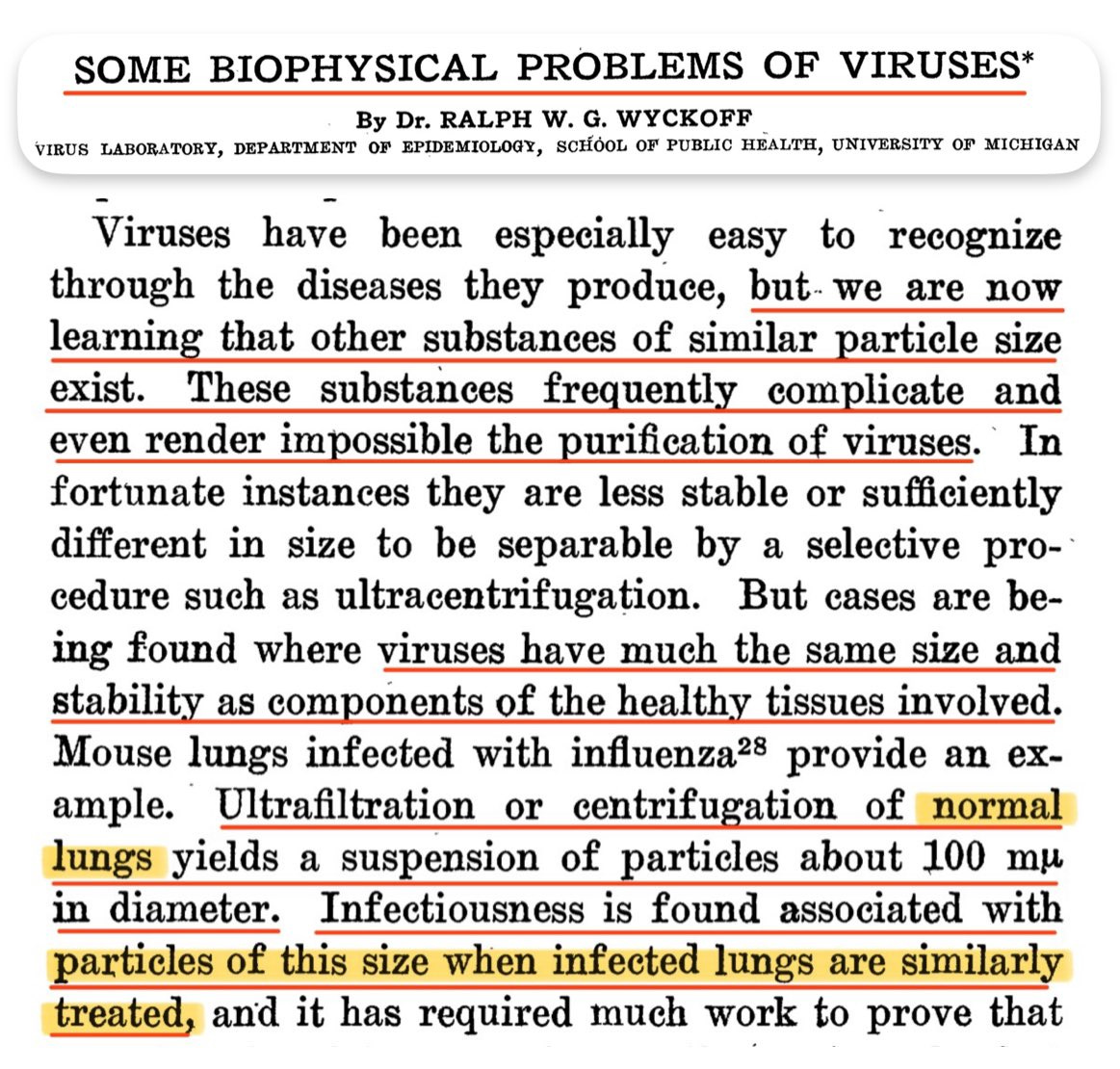
Virologist learned that particles of the same morphology are found in both healthy and diseased tissue, as outlined by a 1945 article entitled Some Biophysical Problems of Viruses.
“Viruses have been especially easy to recognize through the diseases they produce, but we are now learning that other substances of similar particle size exist. These substances frequently complicate and even render impossible the purification of viruses … Cases are being found where viruses have much the same size and stability as components of the healthy tissues involved. Mouse lungs infected with influenza provide an example. Ultrafiltration or centrifugation of normal lungs yields a suspension of particles about 100 mµ in diameter. Infectiousness is found associated with particles of this size when infected lungs are similarly treated.”
The same was described in a 1946 publication.
“The isolation of some animal viruses is complicated by the presence of normal materials which are indistinguishable chemically and physically from the infectious material.”
Virologists discovered that the particles imagined to be viruses were found both in ‘infected’ and ‘uninfected’ material.
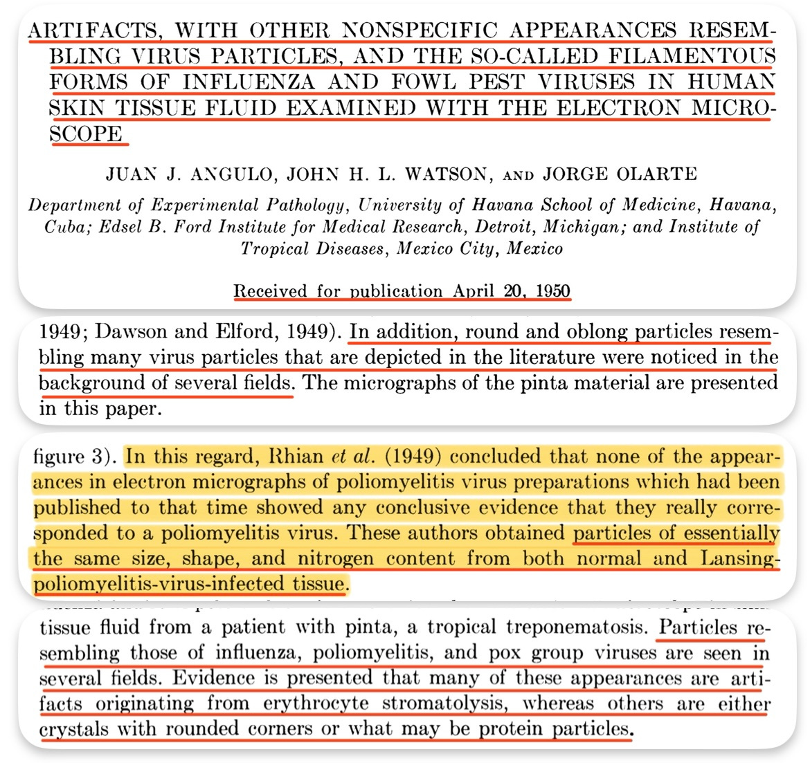
Such was reported in a 1950 study entitled Artifacts, with other nonspecific appearances resembling virus particles.
“Round and oblong particles resembling many virus particles that are depicted in the literature were noticed in the background of several fields.”
[…]
“Particles resembling those of influenza, poliomyelitis, and pox group viruses are seen in several fields. Evidence is presented that many of these appearances are artifacts originating from erythrocyte stromatolysis, whereas others are either crystals with rounded corners or what may be protein particles.”
The authors described the work of a group of virologists who perfomed control experiments and found particles indistinguishable from polio “viruses” in normal uninfected tissue.
“Rhian et al. (1949) concluded that none of the occurrences in electron micrographs of preparations of poliomyelitis virus published up to that time showed any conclusive evidence that they really corresponded to poliomyelitis virus. These authors obtained particles of essentially the same size, shape, and nitrogen content from both normal and Lansing poliomyelitis virus-infected tissue.”
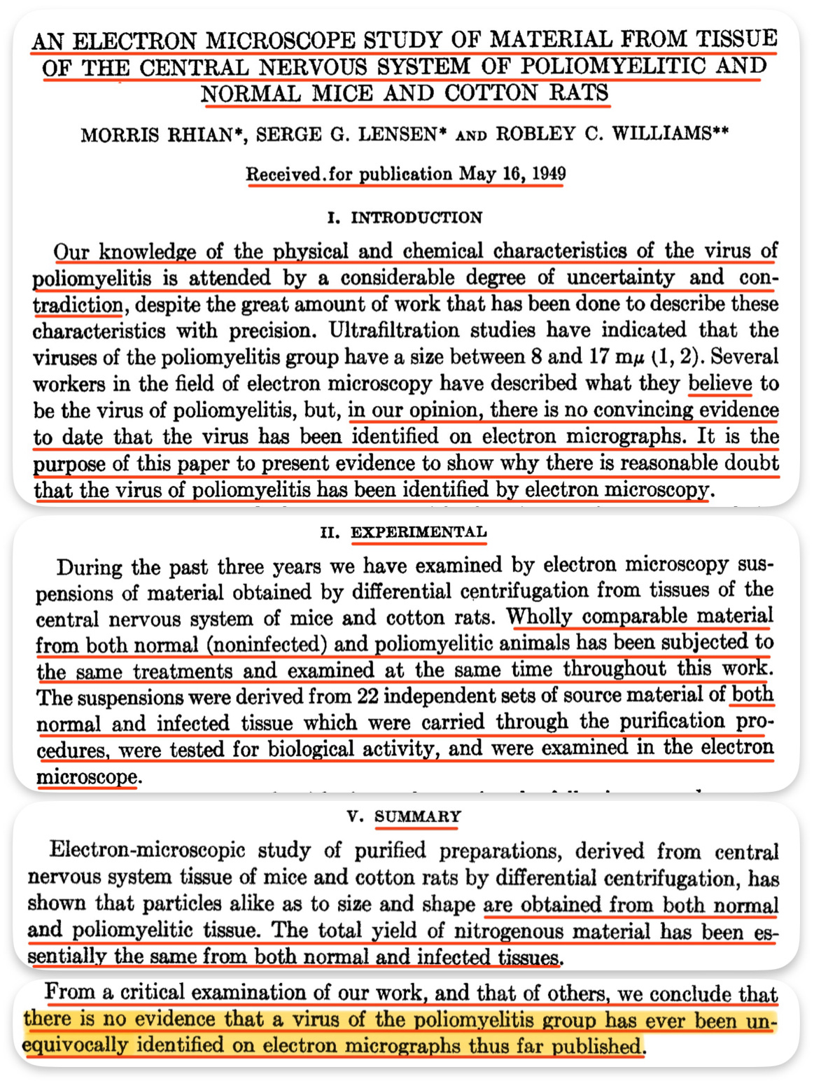
These virologists concluded that the imaged particles purporting to depict polio “viruses” were simply non-specific cellular debris, as described in their 1949 publication entitled An Electron Microscope Study of Material from Tissue of the Central Nervous System of Poliomyelitic and Normal Mice and Cotton Rats.
“Our knowledge of the physical and chemical characteristics of the virus of poliomyelitis is attended by a considerable degree of uncertainty and contradiction, despite the great amount of work that has been done to describe these characteristics with precision. Ultrafiltration studies have indicated that the viruses of the poliomyelitis group have a size between 8 and 17 mµ (1, 2). Several workers in the field of electron microscopy have described what they believe to be the virus of poliomyelitis, but, in our opinion, there is no convincing evidence to date that the virus has been identified on electron micrographs. It is the purpose of this paper to present evidence to show why there is reasonable doubt that the virus of poliomyelitis has been identified by electron microscopy.”
[…]
“During the past three years we have examined by electron microscopy suspensions of material obtained by differential centrifugation from tissues of the central nervous system of mice and cotton rats. Wholly comparable material from both normal (noninfected) and poliomyelitic animals has been subjected to the same treatments and examined at the same time throughout this work. The suspensions were derived from 22 independent sets of source material of both normal and infected tissue which were carried through the purification procedures, were tested for biological activity, and were examined in the electron microscope.”
[…]
“Electron-microscopic study of purified preparations, derived from central nervous system tissue of mice and cotton rats by differential centrifugation, has shown that particles alike as to size and shape are obtained from both normal and poliomyelitic tissue. The total yield of nitrogenous material has been essentially the same from both normal and infected tissues.”
[…]
“From a critical examination of our work, and that of others, we conclude that there is no evidence that a virus of the poliomyelitis group has ever been unequivocally identified on electron micrographs thus far published.”
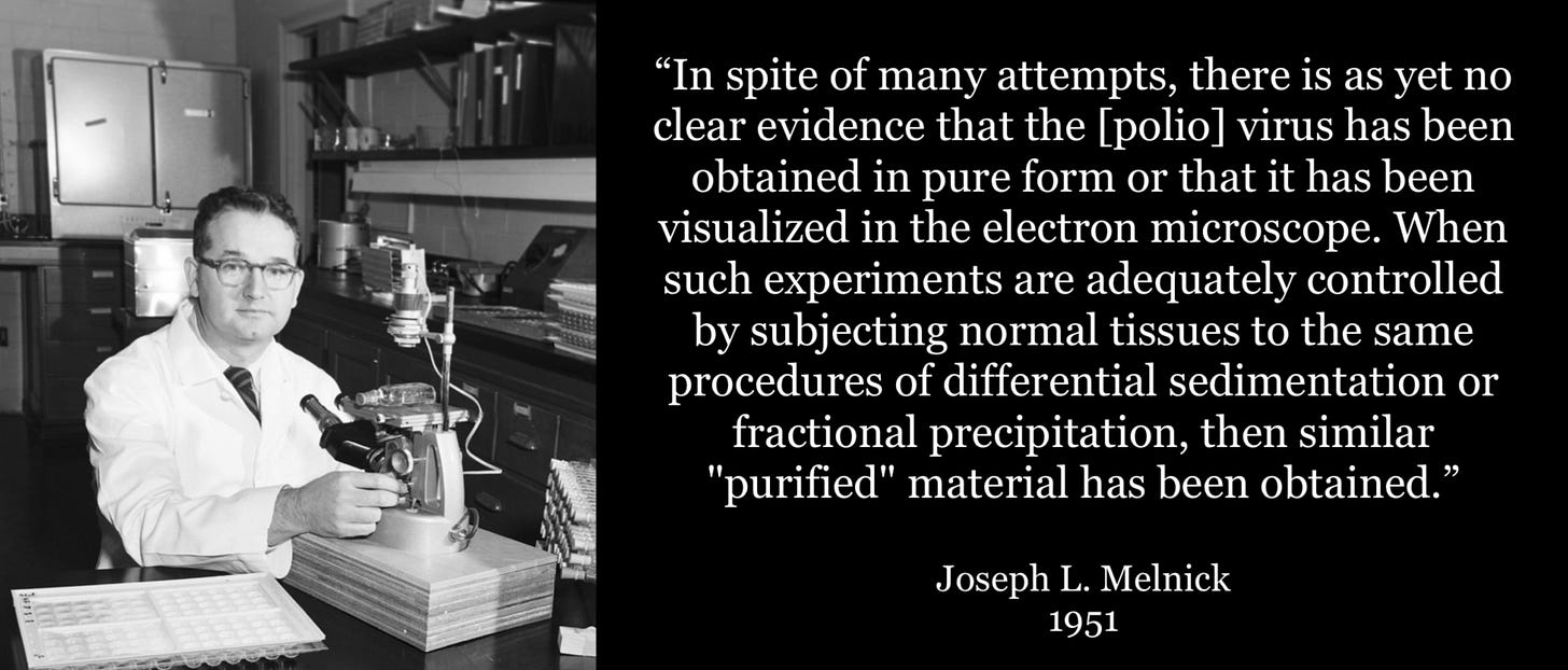
In agreement, virologist Joseph L. Melnick stated in 1951 that there was no clear evidence that the polio virus had been isolated and imaged, because when experiments were adequately controlled by subjecting normal tissues to the same procedures, no difference was found in normal tissue compared to the ‘infected’ tissue.
“In spite of many attempts, there is as yet no clear evidence that the [polio] virus has been obtained in pure form or that it has been visualized in the electron microscope. When such experiments are adequately controlled by subjecting normal tissues to the same procedures of differential sedimentation or fractional precipitation, then similar "purified" material has been obtained (43, 82, 83).”
An electron micrograph from a 1954 publication depicted particles from uninoculated tissue, indistinguishable from “viruses”, allegedly destroying a cell.
Virologist Sven Gard admitted in 1955 that viruses were artifacts.
“Electron microscopy has sometimes been called the science of artefacts. Even if this is a slight exaggeration, production of artefacts is one of the great problems facing the student in this field. As an illustration, it may be sufficient to mention that the virus of Newcastle disease assumes on the electron-microscope screen the shape of spheres, dumb-bells, tailed tadpoles, or short rods, depending upon the salt concentration of the medium from which the specimen is prepared for microscopy. The virus seems to be insoluble in the absence of electrolytes, flocculating in the shape of spherical particles, while in the presence of salts it is soluble, assuming a rod shape.”
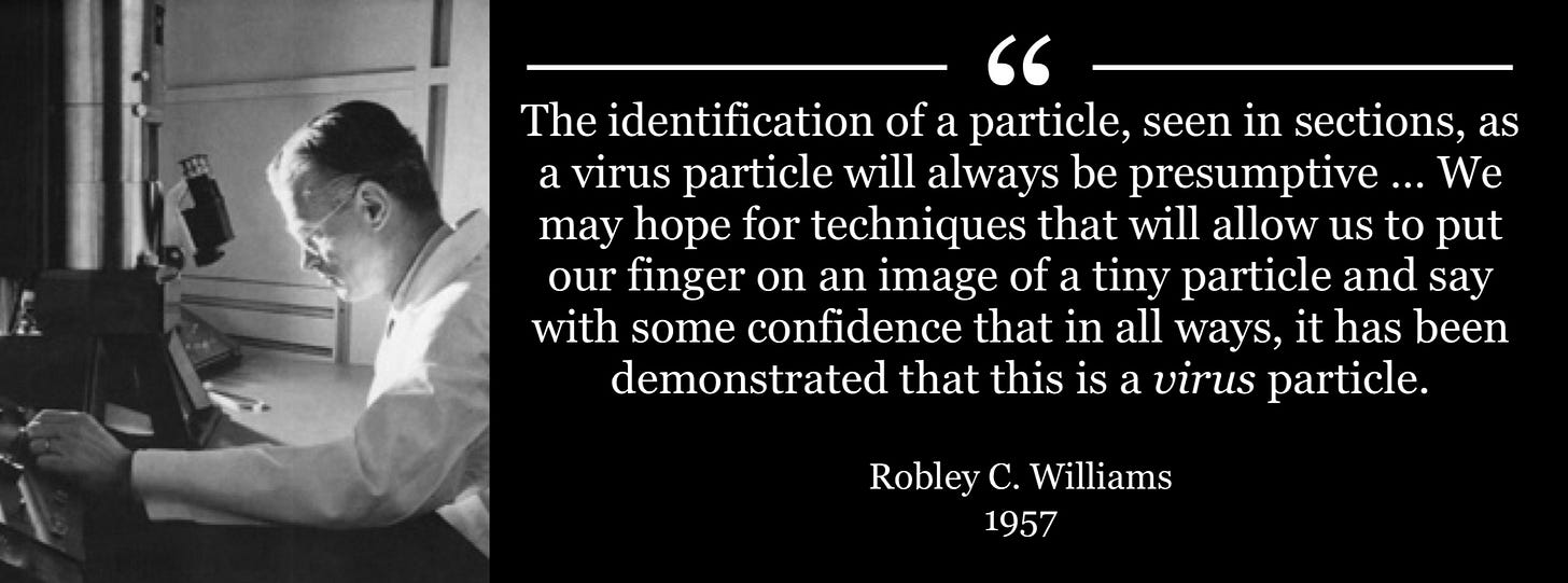
In 1957, virologist Robley C. Williams admitted that there was no certainty that the particles purporting to depict ‘viruses’ actually were viruses.
“The identification of a particle, seen in sections, as a virus particle will always be presumptive … Eventually, also, we may hope for techniques that will allow us to put our finger on an image of a tiny particle and say with some confidence that in all ways, save infectivity, it has been demonstrated that this is a virus particle.”
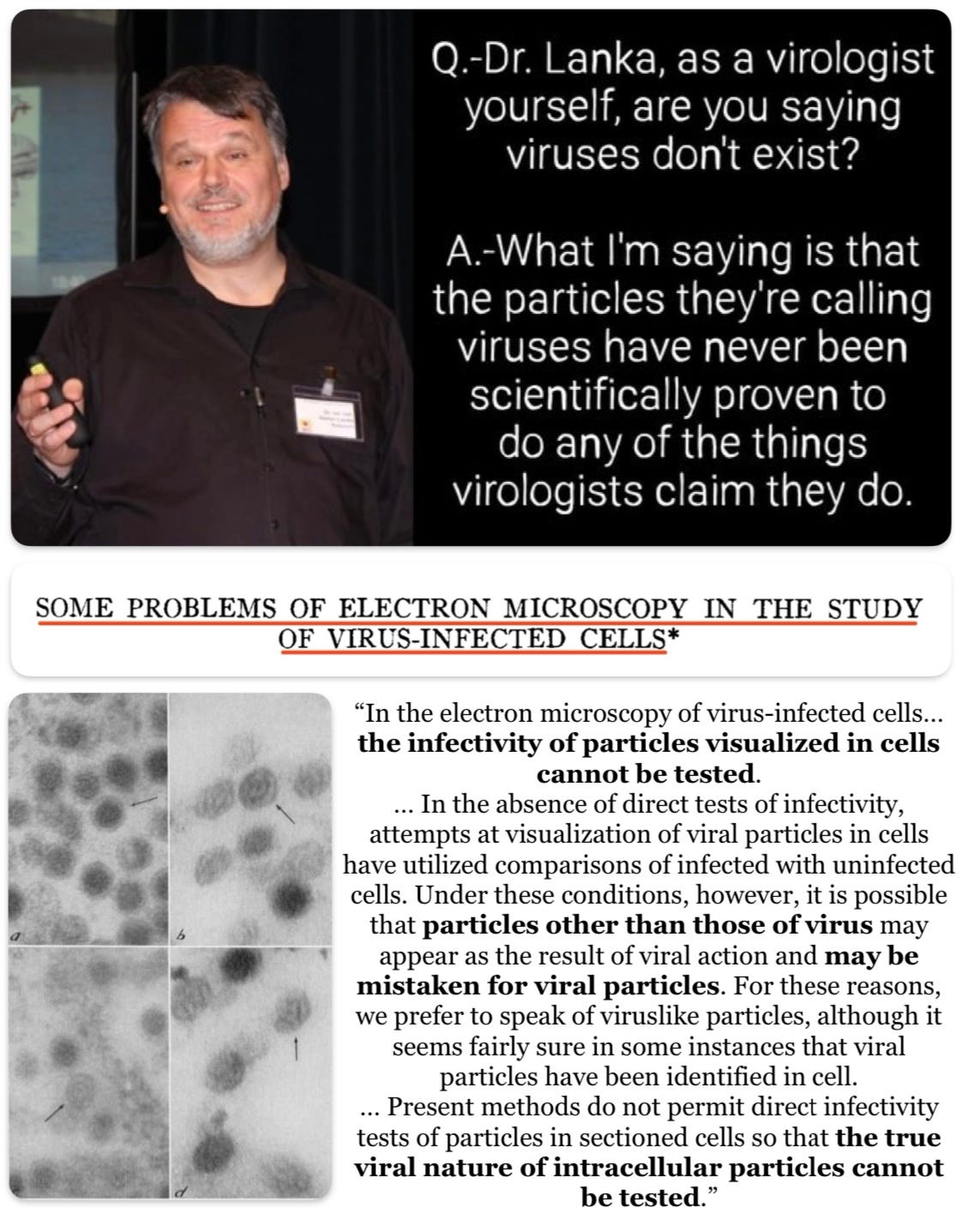
That the biological role of the particles said to be viruses remained undetermined was acknowledged in a 1959 publication entitled Some Problems of Electron Microscopy in the Study of Virus-Infected Cells. The authors recognized that particles may be mistaken for ‘viruses.’
“In the electron microscopy of virus-infected cells... the infectivity of particles visualized in cells cannot be tested.”
[…]
“In the absence of direct tests of infectivity, attempts at visualization of viral particles in cells have utilized comparisons of infected with uninfected cells. Under these conditions, however, it is possible that particles other than those of virus may appear as the result of viral action and may be mistaken for viral particles. For these reasons, we prefer to speak of viruslike particles, although it seems fairly sure in some instances that viral particles have been identified in cells.”
[…]
“Present methods do not permit direct infectivity tests of particles in sectioned cells so that the true viral nature of intracellular particles cannot be tested.”
A 1960 study named Virus Particles in "Normal" Chick Embryos identified particles morphologically indistinguishable from chicken tumor “viruses” in normal healthy chicken tissue.
“However, several studies also revealed that particles similar to those associated with tumors can be found in a rather high percentage of tissues obtained from "normal" fowls, chicks, and chick embryos (3, 5, 6, 15). It was concluded that these were viruses of an unknown variety, but most likely tumor viruses, which were carried within the tissues of apparently "normal" birds. The biological significance of these virus particles found within "normal" tissues is as yet poorly understood. The present report deals with such virus particles in "normal" chick tissues. Particles which morphologically are indistinguishable from chicken tumor viruses were found incidentally in the course of cytologic studies of chick embryo aorta and liver.”
[…]
“The work of a group of investigators in Paris has shown that virus-like particles morphologically indistinguishable from the known chicken tumor viruses can also be found in thin sections of tissues obtained from "normal" chickens, chicks, or chick embryos (3, 5). Similar particles were seen in sections of tissue cultures originating from "normal" chick tissues (15). The present report confirms this previous information.”
[…]
“Of major interest is, we believe, the fact that the cells of the "infected" tissues as described here looked entirely "normal".”
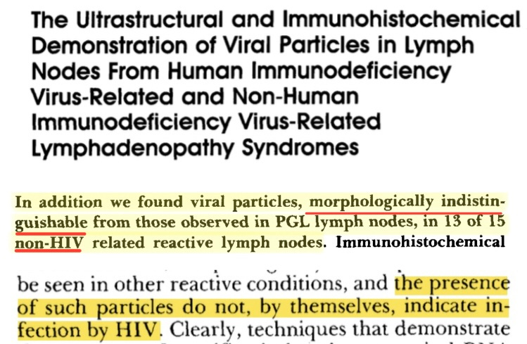
During the 1980s, particles identified as HIV were observed in tissue of AIDS patients. However, identical particles were also observed with the same frequency in tissue of patients who did not have AIDS and were not at risk of AIDS.
Such was outlined in an extensive, detailed and blinded electron microscopic study from Harvard. ‘HIV particles’ were found in 18/20 (90%) of patients with ‘AIDS’ whereas identical particles were found in 13/15 (87%) of patients without ‘AIDS.’
As electron microscopy has improved, microbiologists still find indistinguishable particles in uninfected tissue. A 2020 paper found particles identical to “SARS-CoV-2” in tissues from patients negative for covid as well as in biopsies from the pre-covid era.
“Since the discovery of the causative agent for the novel severe acute respiratory syndrome (SARS)–like pneumonia syndrome pandemic that started in China in 2019 (1,2), a coronavirus named SARS coronavirus 2 (SARS-CoV-2), electron microscopy images have populated the medical literature (2) and media outlets alike displaying the characteristic 60–140 nm round particles surrounded by a “corona” of 9–12 nm distinctive spikes (2). Although many of these images were obtained after “in vitro” infection of cultured cells with SARS-CoV-2 (2) and are thus likely a true representation of viral particles, we have observed morphologically indistinguishable inclusions within podocytes and tubular epithelial cells both in patients negative for coronavirus disease 2019 (COVID-19) as well as in renal biopsies from the pre–COVID-19 era.”
Additional information about this topic can be found on Category: Electron Microscope Images at viroliegy.com.
Futher reading on the concept and history of viruses can be found in the article below.








You should do a writeup on rabies at some point. The entire story is ridiculous, from the extremely complex behavior of "the virus" (<12,000 nucleotides in an RNA virus that can infect and spread throughout your body into your brain where it lies in wait to cause neurological problems before migrating into your mouth to infect your saliva and inducing you to bite people to "spread it" - complex behavior considering saliva and mucrous consists of at least 12 mucinic compounds encoded each by genes of 7,000 nucleotides or more), its purported lethal fatality rate (100% in humans!), the exceptionally few - near zero - "rabies deaths" over the past 100 years (in the US anyway), the "vaccines" over the years (initially injecting you or your dog with goat or rabbit brains - which caused autoimmune neural problems, sadly), the fact that it caused near-zero problems until the 1980s when they started of course pushing new "vaccines", it's "isolation" and "vaccine" development by Pasteur in 1885 (rabbit brains injected into living rabbit brains ground and injected into yet more poor rabbits' brains), the notion that (despite the virus TEAMING in an animal's saliva) the only way to "detect" it is to kill the animal and do a dFA test on its brain tissue, and so on. The entire story is insane and ridiculously silly. But when you think about it, really mostly just psycho-killer-Jeffrey-Dahmer-animal-torturing level insane given certain government mandates on the rabies vaccines in different locales and the need to slice and dice brains to "find it."
And, to boot, in many states, all pets or people who work in "high risk" groups have to get vaccinated every 3 years. Why? Because you simply *MUST* have "rabies" "antibodies" floating around in your body at all times to "be immune." Other vaccines can "protect you" for 10 years, sometimes forever, but not rabies, it's 1-3 years max "because antibodies".
COVID was a real eye-opener into the total lunacy and insanity of virology and vaccines, I gotta say. Anyhow thank's for all the other articles you've written each deep dive is a learning experience into how flimsy this "science" is.
Great work like always!